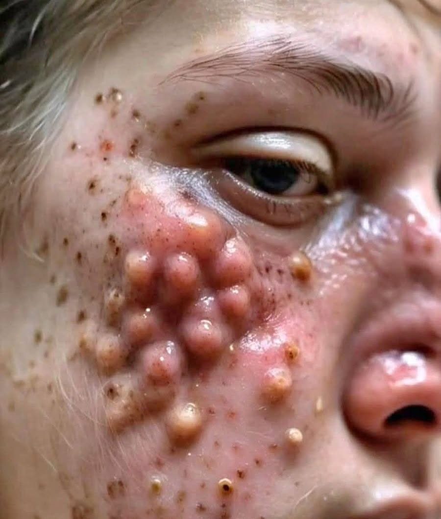
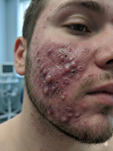
The image portrays an intense, highly localized clusters of atypical dermatological abnormalities on the central facial region. Visually, this is characterized by a dense grid-like pattern of highly elevated, firm, and fluid-filled nodulocystic structures. Surrounding and interspersed among these intensely inflamed lesions are a multitude of deeply dilated, open follicular openings with stark, dark centers that simulate macro-comedones (large blackheads). The underlying skin structure exhibits severe erythema (redness) and acute localized swelling, indicating an overwhelming inflammatory process beneath the surface.
In a clinical context, a real presentation resembling this would represent an extreme progression of nodulocystic acne or acne conglobata. This happens when individual, deep-seated blockages do not simply rupture and heal, but instead expand horizontally and vertically within the lower dermal layers. As multiple adjacent oil glands and hair follicles rupture below the surface, they form interconnected tracts or “tunnels” of active infection and inflammation, creating a unified, painful mass that significantly alters the outer contour of the skin.
Why It Happens: The Multi-Step Biological Trigger
The physiological development of deep, cystic skin lesions of this scale relies on a cascade of events taking place within the pilosebaceous unit (the hair follicle and its associated oil gland). This process is typically driven by four primary overlapping mechanisms:
- Severe Seborrhea (Oil Overproduction): Triggered heavily by genetic predispositions or systemic hormonal surges, the sebaceous glands produce an excessive volume of heavy sebum.
- Follicular Hyperkeratinization: The cells lining the inside of the pore shed abnormally and rapidly. Instead of rising to the surface and washing away, these sticky, dead cells clump together with the excess oil, forming a dense, unyielding plug that completely seals the pore.
- Microbial Overgrowth: Deep within the oxygen-deprived environment of the blocked pore, Cutibacterium acnes bacteria multiply exponentially, feeding on the trapped sebum.
- Extreme Immune Cascade: The body recognizes the ballooning pore and bacterial proliferation as an emergency. It dispatches a massive wave of white blood cells to attack the area. When the walls of the follicle finally rupture under pressure, this bacteria and cellular debris leak directly into the surrounding deep dermis, triggering a violent, painful inflammatory response that forms large, physical nodules.
How to Heal: Comprehensive Dermatological Interventions
Resolving severe, structural skin inflammation requires intensive systemic medical management supervised by a specialist. Standard over-the-counter washes, topical scrubs, or physical extraction attempts are entirely ineffective for deep-seated lesions and will drastically worsen inflammation, cause secondary infections, and guarantee severe permanent scarring.
Healing is achieved through a multi-pronged clinical strategy. The core baseline treatment for chronic, nodulocystic presentations is oral retinoid therapy, such as isotretinoin. This systemic medication temporarily reshapes the skin’s biology by aggressively shrinking the sebaceous glands, reducing oil production, normalizing the lifecycle of skin cells to prevent pore blockages, and heavily calming the systemic inflammatory response.
To manage immediate pain and halt active tissue destruction, dermatologists utilize short-term targeted interventions. This can include short courses of oral antibiotics or anti-inflammatory medications to decrease the immediate bacterial and white blood cell load. Additionally, specialists may perform localized intralesional corticosteroid injections directly into the center of the largest nodules. This delivers a highly concentrated anti-inflammatory agent precisely where it is needed, rapidly collapsing the swelling within hours, alleviating intense physical pressure, and safeguarding the surrounding skin architecture from permanent, deep scarring.
Important Note on Media Authenticity: While the text above details the true medical science behind severe nodulocystic conditions, it is important to analyze the media source itself. This image bears classic hallmarks of an AI-generated filter or digital manipulation designed specifically to look shocking and trigger “trypophobia” (a fear or aversion to clusters of small holes or bumps). Real clinical acne conglobata or severe cystic acne presents with irregular shapes, asymmetrical swelling, varying stages of healing, and fluid dynamics that do not conform to the highly uniform, neatly repeating grid pattern seen across the cheek in this picture. Online advertisements or social media posts frequently use these exaggerated, digitally synthesized images alongside a “Swipe Up” call-to-action to shock viewers into clicking links or purchasing unrelated skincare products.
- Medical Disclaimer: This analysis is provided strictly for educational and informational purposes based on the visual features of the provided media. It is not a substitute for professional medical evaluation, diagnosis, or treatment. Always consult a licensed dermatologist or medical professional regarding actual severe skin conditions.*